《医学影像学英语教程》曲晓峰,边杰,郭冬梅主编|(epub+azw3+mobi+pdf)电子书下载
图书名称:《医学影像学英语教程》
- 【作 者】曲晓峰,边杰,郭冬梅主编
- 【页 数】 468
- 【出版社】 北京:中国协和医科大学出版社 , 2015.07
- 【ISBN号】978-7-5679-0221-3
- 【分 类】医学摄影-英语-教材
- 【参考文献】 曲晓峰,边杰,郭冬梅主编. 医学影像学英语教程. 北京:中国协和医科大学出版社, 2015.07.
图书封面:
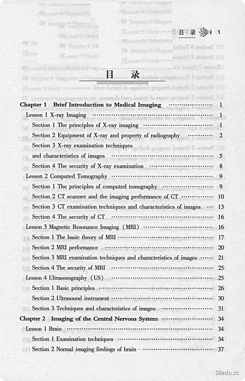
图书目录:


《医学影像学英语教程》内容提要:
本书是医学影像学英语教程。
《医学影像学英语教程》内容试读
Chapter 1 Brief Introduction to Medical Imaging1
n blles as dou lab-tbM
isnsb-wol oui-visnsh daid035w10
Chapter 1
Brief Introduction to Medical Imaging
Lesson 1 X-ray Imaging
X-ray imaging has been applied in clinical settings to help physicians diag-nose diseases for over one hundred years.Today it is still an important and irre-placeable imaging technique.
Section 1 The principles of X-ray imaging
X-ray can make the structures inside human body visible on fluorescentscreens or films,which is attributed to both the properties of X-ray (namely,itspenetrating,fluorescent,and sensitization effects)and the differences indensity and thickness among different human tissues.When X-ray penetrates dif-ferent tissues and structures with varying X-ray absorption degrees inside humanbody,there exist differences in the amount of X-ray photons that reach the fluo-rescent screens or films.Furthermore,because of the fluorescence effect andsensitization effect,the differences in X-ray absorption can produce shades ofgray that contain information on radiography or fluoroscopy.
The human body is composed of different materials containing different ele-ments that absorb X-ray in varying degrees.Based on the density,the tissues in-side human body can be divided into three categories:a)high-density tissuessuch as bones and calcification;b)moderate-density tissues such as cartilages,muscles,nerves,parenchymal organs,connective tissues,and body fluid;and c)low-density tissues such as fatty tissues,respiratory tract,gastrointestinal tract,paranasal sinus,and mastoid sinus.High-density tissuessuch as bone absorb more X-ray and allow less X-ray to penetrate,thus presen-ting white on X-ray films.In contrast,low-density materials such as lung with
2医学影像学英语教程d【。
air appear black.Moderate-density tissues such as solid organ appear graybetween high density-tissues and low-density tissues Fig 1-1-1 )Therelationship between X-ray absorption and thickness is intuitively obvious:athick piece of any material absorbs more X-ray than a thin one;thus,the degreeof black-white image on X-ray films is related with the density of organizationalstructures and affected by their thickness.
t10889.
-yaib sneiniggrg qlad a
ed anrgnmi wr-X
-omu bna msroqmi nt rg
avo 1ol as8nskih snoyl gnigs eldasai
pnipsmi v
noitoee
ono sldiiv
m0的-
aitm)X lo
idw andn to nnoa
ni woorsslih on br
errgDanitdgnng
dnng (aFig 1-1-1 Normal chest:P-A positionlhi山sit话
nacud abiani eamasb mou
s1时oabg≌U2过bs
ouds山sss鲤e anoforig-lo innoms o山di soommofib Jai9sod,bod
Diseases can change tissue density in human body.When this changereaches a certain degree,it can make the normal black-white image change in X-ray films.Therefore,pathological changes with different tissue density can pro-duce corresponding pathological X-ray images.eoqinoo ai tbod namd s
orasb arivy ni yei-K cioads lodt sloam
的如Section 2 Equipment of X-ray and property
of radiography
:bisll yhod brm Bouwit
Tg,90,地n
X-ray examination has been constantly improved with the development of sci-ence and technology.In particular,the past three decades have witnessed thedigitization of X-ray imaging.Nowadays X-ray examination has more diverse ap-
Chapter 1 Brief Introduction to Medical Imaging3
plications.mudni
1 Conventional X-ray and imaging performance
Conventional X-ray includes general radiography,gastrointestinal machine,angiography,mobile X-ray,mammography,and dental radiography,duringwhich the X-ray penetrates the human body and displays the findings on films.Ithas many advantages:a)high spatial resolution of image;b)showing thewhole structure tissue in a wide range;c)low X-ray radiation;and d)eco-nomically affordable.However,it also has many limitations:a)strict radio-graphical conditions;b)low density resolution of image;c)organizationalstructures overlapping each other in the image,which has some impact on displa-ying the pathological change;d)the black-white degree of images is connectedwith the radiographical condition,therefore makes it difficult to display tissues ofdifferent densities at the same time;and e)inconvenient to utilize and managethe X-ray films.
ortnlm-e
gd yool bns sq
2 Digital X-ray equipment and X-ray properties
Based on technical principles,digital X-Ray equipment includes computedradiography (CR),which can be combined with conventional equipment,anddigital radiography DR )which includes universal,gastrointestinal,mammary,and mobile machines.
However,when taking pictures,X-ray penetrating human body must bepixelized and digitized before computer processing.Unlike CR,which usesimage plates (IPs)as carrier for information of X-ray penetrating human bodyinstead of films,DR uses flat panel detectors.The advantages of digital radio-graphy are as follows:a)the requirements of taking pictures are not limited,thus reducing X-ray radiation dose;b)it improves the quality of images;c)ithas many image post-processing functions such as measurement,edgesharpening,and subtraction;d)the digital data can be printed into films or bevisible on the monitor screens,or be saved in computers.The disadvantages ofCR include:a)its slow imaging makes X-ray examination impossible;and b)it is not highly efficient.However,DR can not only greatly shorten the time of
4藏医学影像学英语教程d【c
imaging in X-ray examinations,but also further improve efficiency,thereby re-ducing exposing dose;DR can perform more image processing,such as volumerendering (multi-plane tomographic image of any thickness in the projected posi-tion can be obtained by performing one scan),automatic mosaic (large-scalesuch as full spine DR seamless image after mosaic can be achieved by one scan)and so on.
3 Digital subtractive angiography system and X-rayimaging performance
Digital subtractive angiography (DSA)system is a combination of computertechnology and conventional angiography.The image acquisition system of DSAwas composed of X-ray image intensifier and high-resolution video camera at first.
Since the framework of DSA looks like a "C",it is also known as C-arm X-raymachine.DSA can be divided into single C-arm and double C-arm or suspensiontype and floor type according to the installation method.There are also mobileDSA and fixed DSA in hybrid operation rooms.
DSA can be used in cardiovascular system imaging and interventionaltherapy.In the past,imaging for the blood vessels was extremely difficult sincethey were surrounded by bones and soft tissues.Now DSA can display cardiovas-cular system more clearly and completely.Among the digital subtractionmethods,the most common algorithm using digital radiographic systems is tempo-ral subtraction method,which can provide legible blood vessel images withoutbeing affected by bone tissues and soft tissues by subtracting the radiographic im-age obtained without a contrast agent from an image taken with a contrast agent bycomputer-assisted techniques (Fig 1-1-2).Blood vessels of 200um or above indiameter can be obtained clearly by this method.So far,DSA is still a goldenstandard in diagnosing cardiovascular diseases as well as an indispensableimaging approach in endovascular interventional treatment.
A.mask image;B.angiogram;C.DSA image.After pixel and digitaltransformation,the digital subtraction of both mask image (A)and angiogram(B),the digitals of bones and soft tissues offset and only intravascular contrastmedia digital remains.After digital/analog conversion,finally we will obtain a
Chapter1 Brief Introduction to Medical Imaging5
Fig 1-1-2 The principle of DSA
u宽3皆
digital subtraction vascular image (C)
Section 3 X-ray examination techniques
and characteristics of images
The differences in the density and thickness of human tissues are the basisfor producing X-ray image contrast,namely natural contrast.Plain film is an im-age that depends on this natural contrast.As for tissues or organs lacking naturalcontrast,higher or lower contrast media are deliberately introduced into thesestructures to produce contrast between them,which is called artificial contrast.
The examination by means of artificial contrast is called X-ray contrast examina-tion.
4
1 Conventional examinations
(1)Radiography
Radiography is applied broadly in checking various parts of human body.
Generally speaking,it is necessary to take two different orientations such as nor-motopia and mediolateral in order to figure the lesion out better and display itsfeature and location.For instance,it can display angulation displacementfracture of one bone in one orientation,which can not be shown in another orien-
6医学影像学英语教程d1的
tation.
(2)Fluoroscopy
At present,FPD and image enhancement television system are twoimportant approaches that are widely applied in gastrointestinal barium imaging,interventional therapy,and reduction of fracture and so on.
2 Special examinations
(1)Soft ray radiography It is used for the examination of soft tissues (es-pecially the breast)based on molybdenum target or rhodium target.
(2)X-ray subtraction technique It can provide pure soft tissue image orbone tissue image with CR or DR subtraction function.For example,one sub-traction chest pure soft image will enhance the positive proportion of thediagnostic results for tiny non-calcified pulmonary nodules.
(3)Volume tomography
8 nongse
With this DR technique,we can obtain multi-dimension images in anydepth and thickness.For example,it is possible for us to figure out the bone de-struction through observing the continuous structures of centrums and vertebral ar-ches when checking vertebral column,which is difficult to display in plain films.
3 X-ray angiography
(1)The types and applications of contrast agents
a)medicinal barium sulfate,just used in esophagus and gastrointestinaltract contrast radiography;and b)water-soluble organic iodine contrast agent:ionic and non-ionic,mainly used in angiography,intravascular interventionaltherapy,urography,hystero-salpingography,contrast fistulography,and T-tubecholangiography
Water-soluble organic iodine contrast agent injected into the vessel maycause adverse reactions,which can be severe in some cases.The pervasive ap-plication of contrast agent is non-ionic for less and light degree of adverse reac-tions,whereas ionic contrast agent has been obsolete.Besides,water-solubleorgan iodine contrast agent should be used with caution (or even forbidden)in
···试读结束···
作者:柳建华
链接:https://www.58edu.cc/article/1566957699382431745.html
文章版权归作者所有,58edu信息发布平台,仅提供信息存储空间服务,接受投稿是出于传递更多信息、供广大网友交流学习之目的。如有侵权。联系站长删除。